
Laboratory gallery
These images show the class of work — microscopy, diffraction, spectroscopy, electrochemistry, powders and a bench laboratory. They are not photographs of NS&TD instruments and do not imply a model or specification.
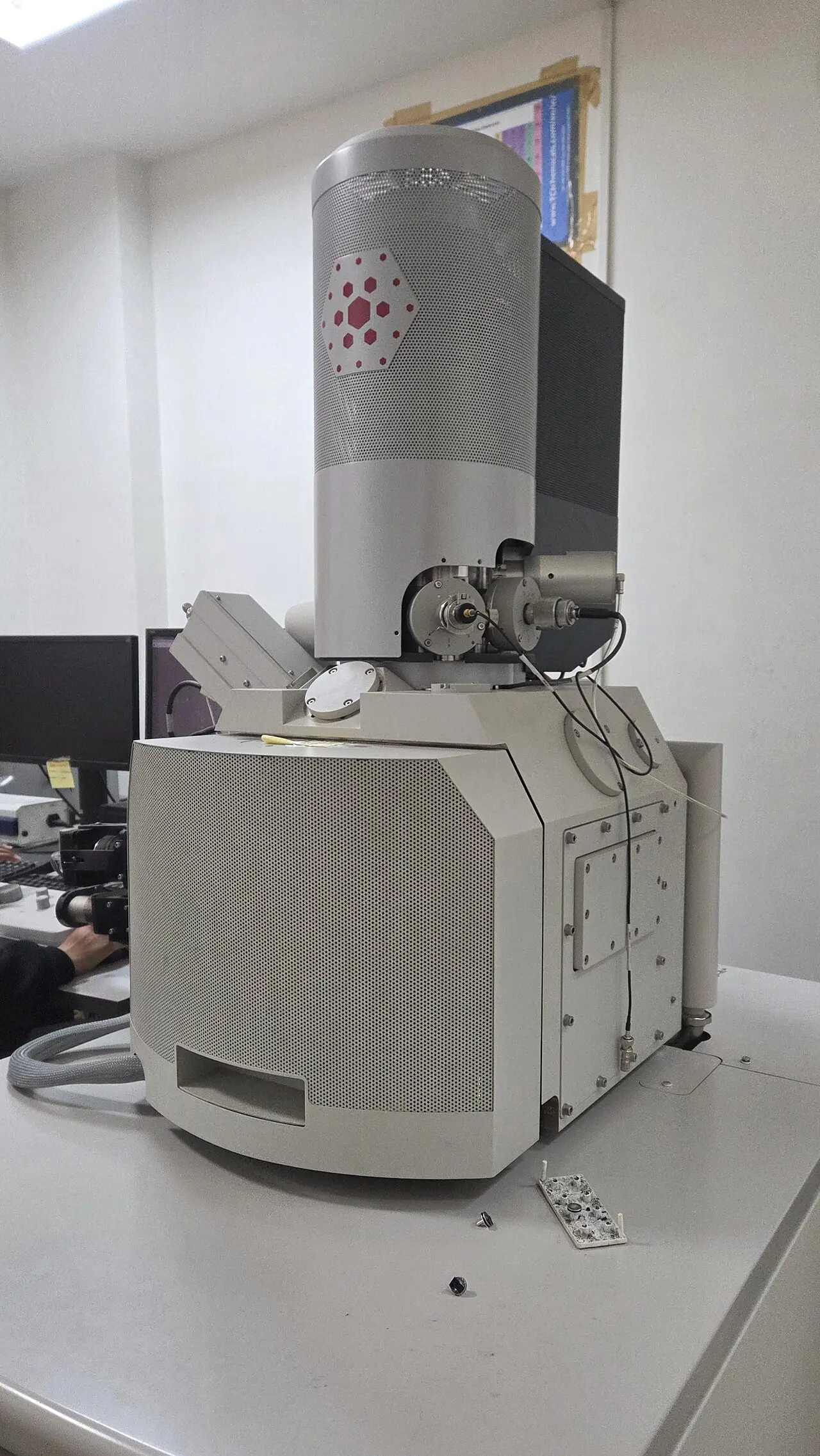
Field-emission SEM is the facility’s surface-imaging technique, used with EDX for elemental composition.
Gannu03, CC BY-SA 4.0. Source

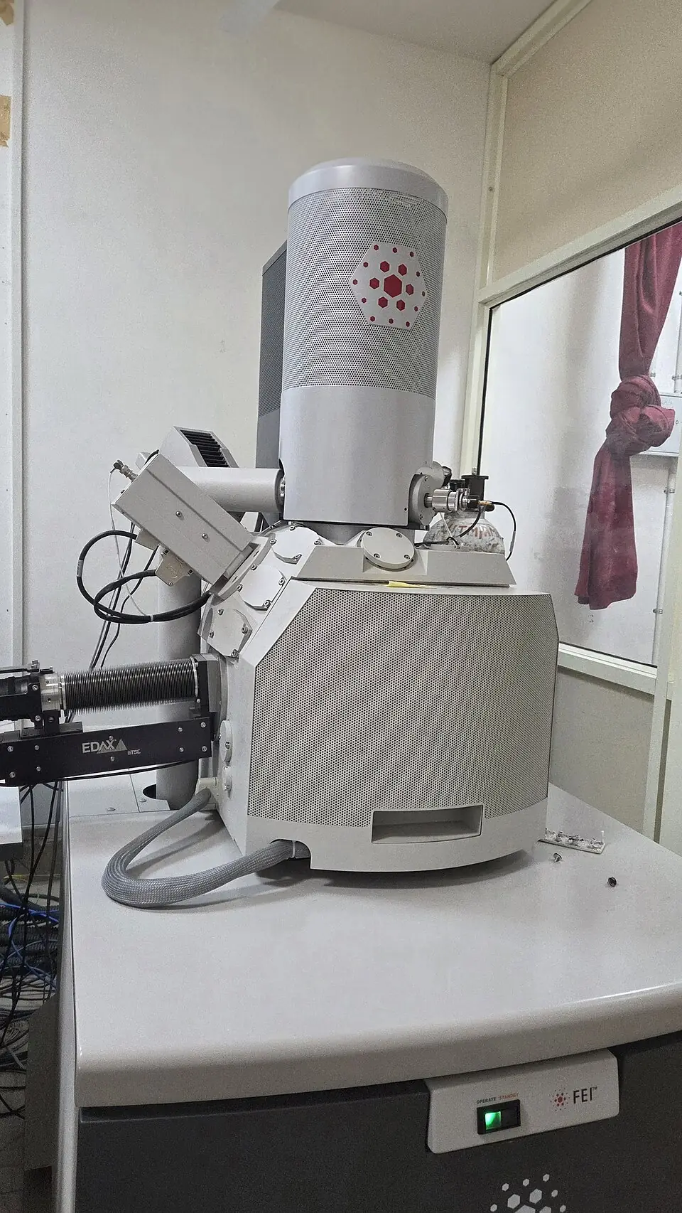
Close view of the electron column and EDX detector port. Illustrative of microscopy, not an NS&TD instrument.
Gannu03, CC BY-SA 4.0. Source

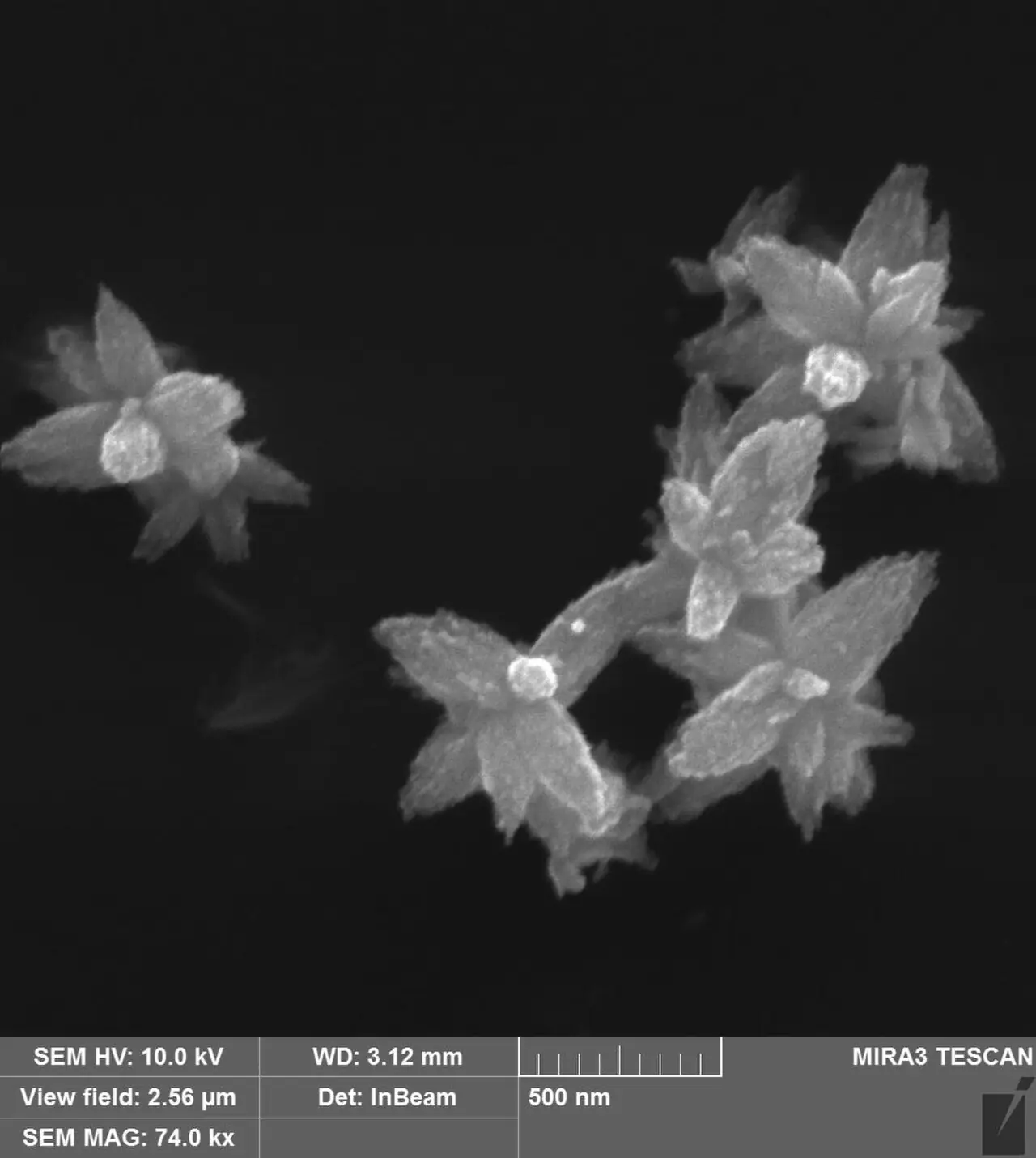
Electron micrograph of zinc oxide nanoflowers, standing in for nanoscale surface imaging.
Oleksandr Smirnov, CC BY 4.0. Source

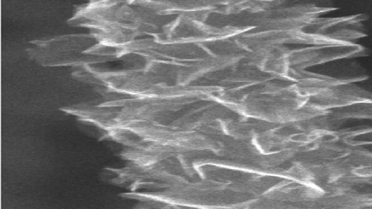
Graphene foliates on a carbon nanotube. Used for the carbon-materials research area.
BRStoner, Duke University, 2011, CC BY-SA 3.0. Source
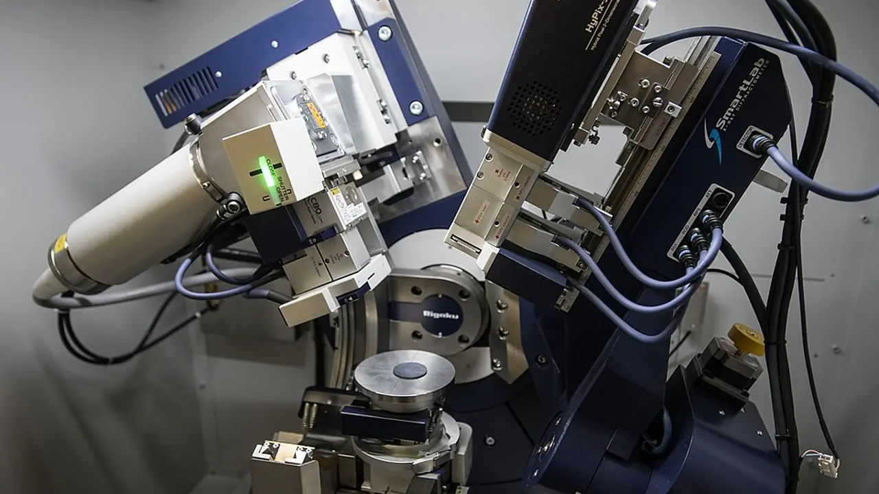
XRD identifies crystalline structure, phase, and crystallite size.
René Volfík, CC BY-SA 4.0. Source

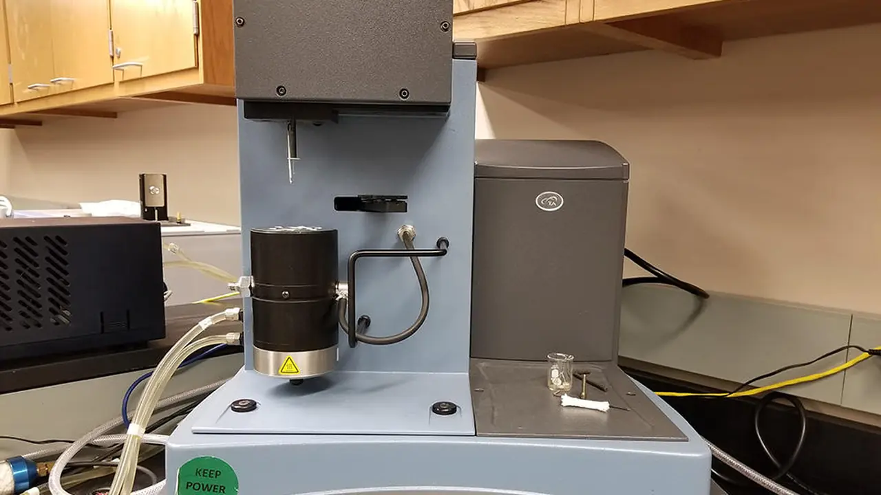
TGA follows mass change with temperature. The facility also lists DSC in this group.
University of Washington MSE laboratory equipment page. Source
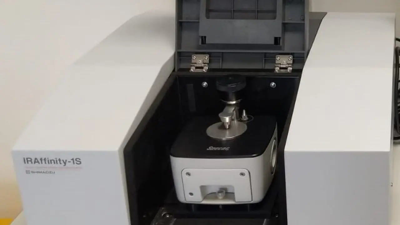
FTIR identifies functional groups from infrared absorption.
Keshavana, CC BY-SA 4.0. Source

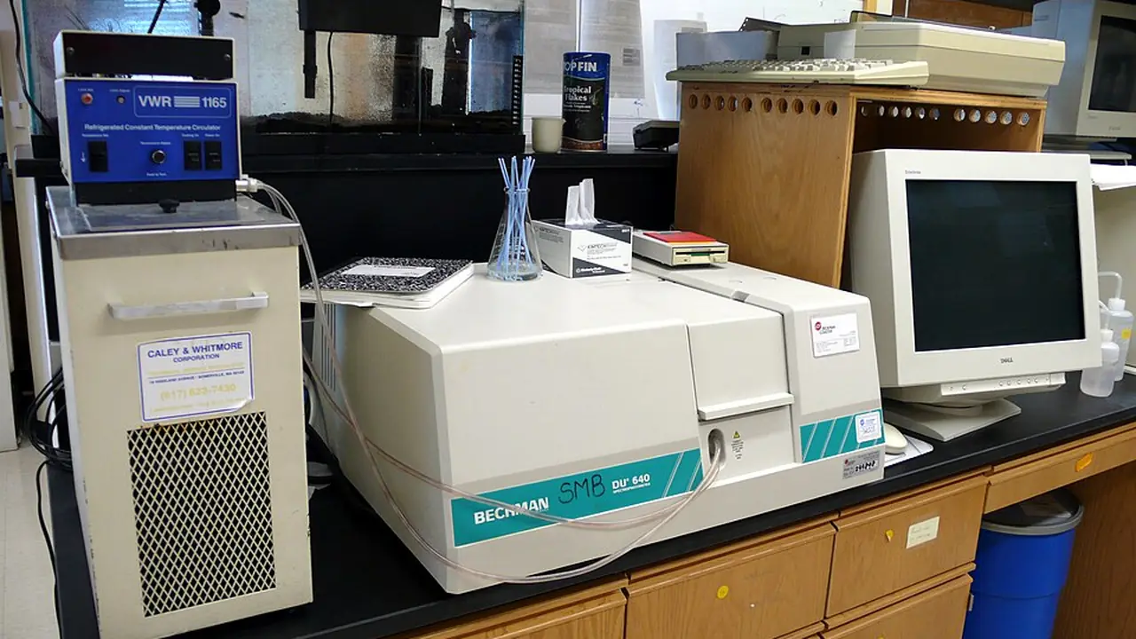
UV-Vis and DRS cover solution and solid-state optical absorption.
Tim Vickers, public domain. Source

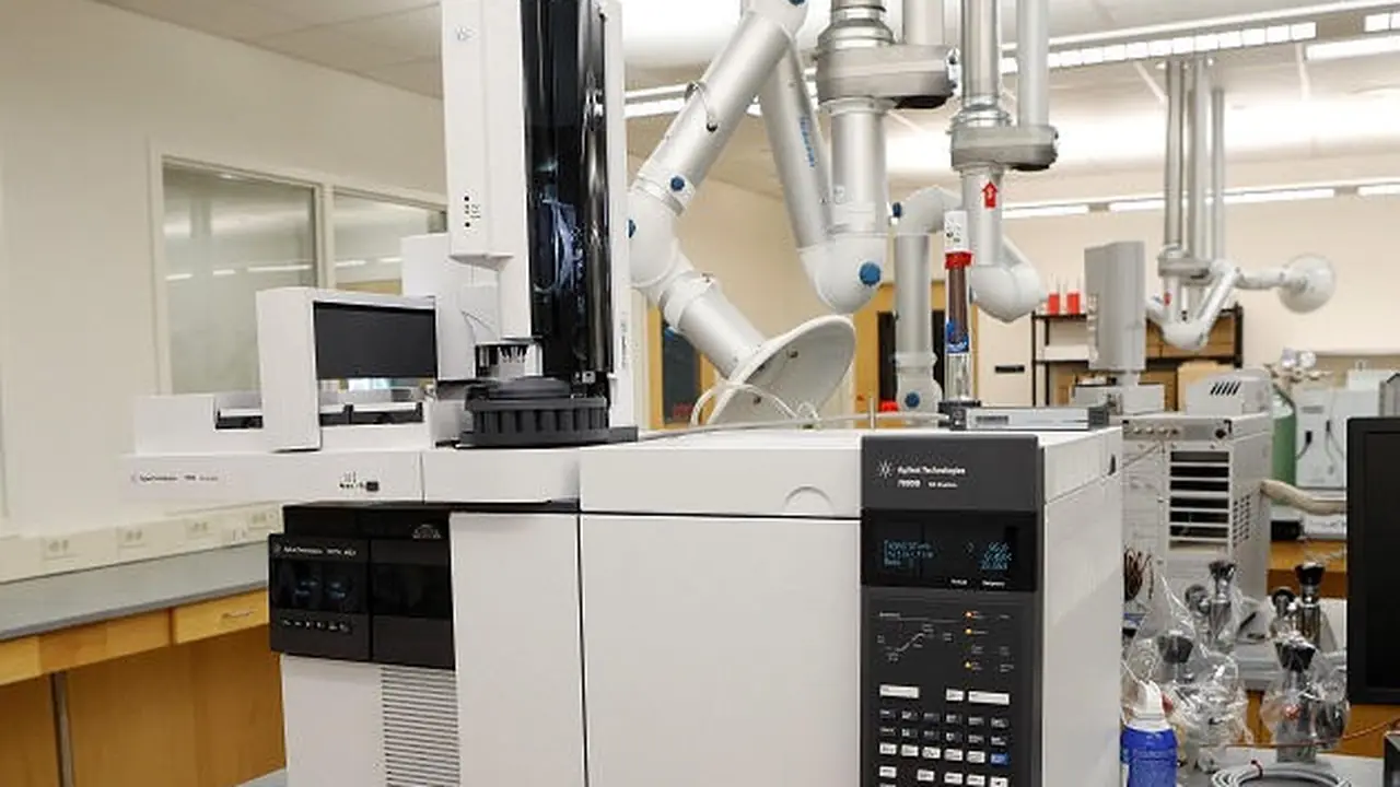
Gas chromatography–mass spectrometry separates and identifies volatile compounds.
Vermont Forensic Laboratory. Source
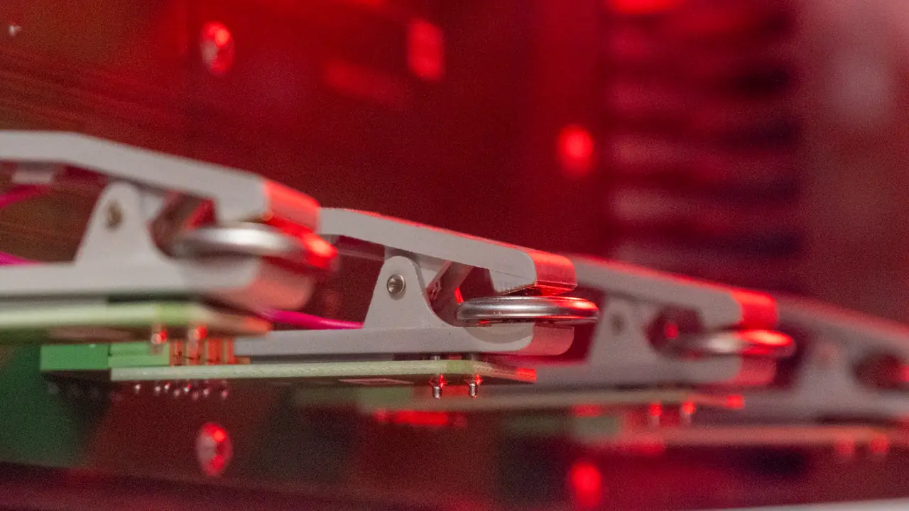
Coin cells in a test fixture, used for the energy-storage area and electrochemical techniques.
Wikimedia Commons. Source

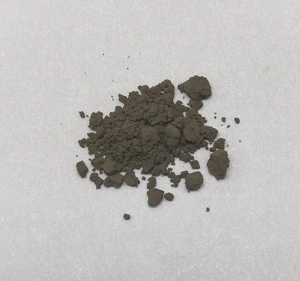
A carbon powder sample. Carbon materials and nanocomposites are named research areas.
Leiem, CC0. Source

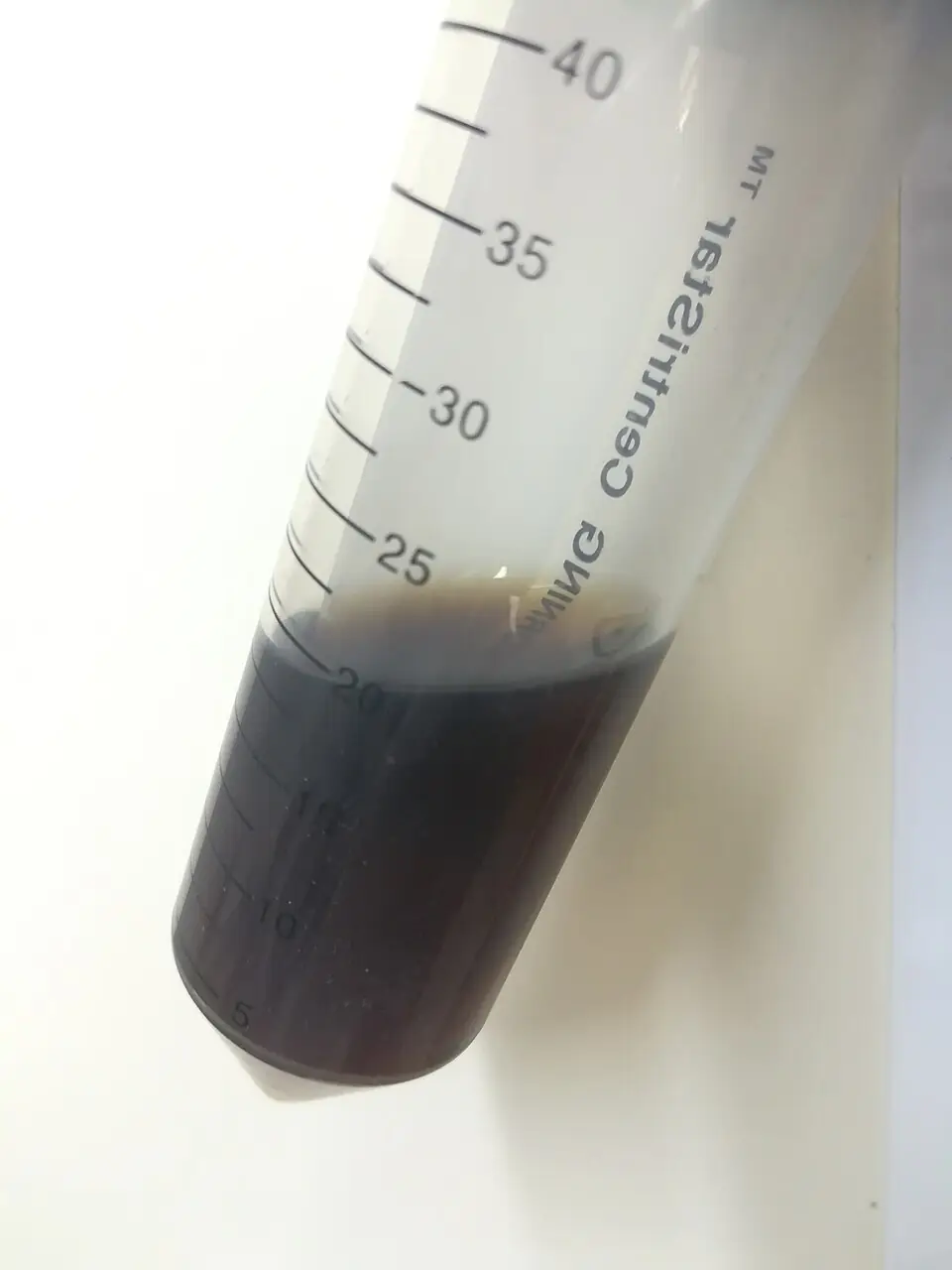
A water dispersion, relevant to zeta-potential samples, which must be liquid.
Leiem, CC0. Source

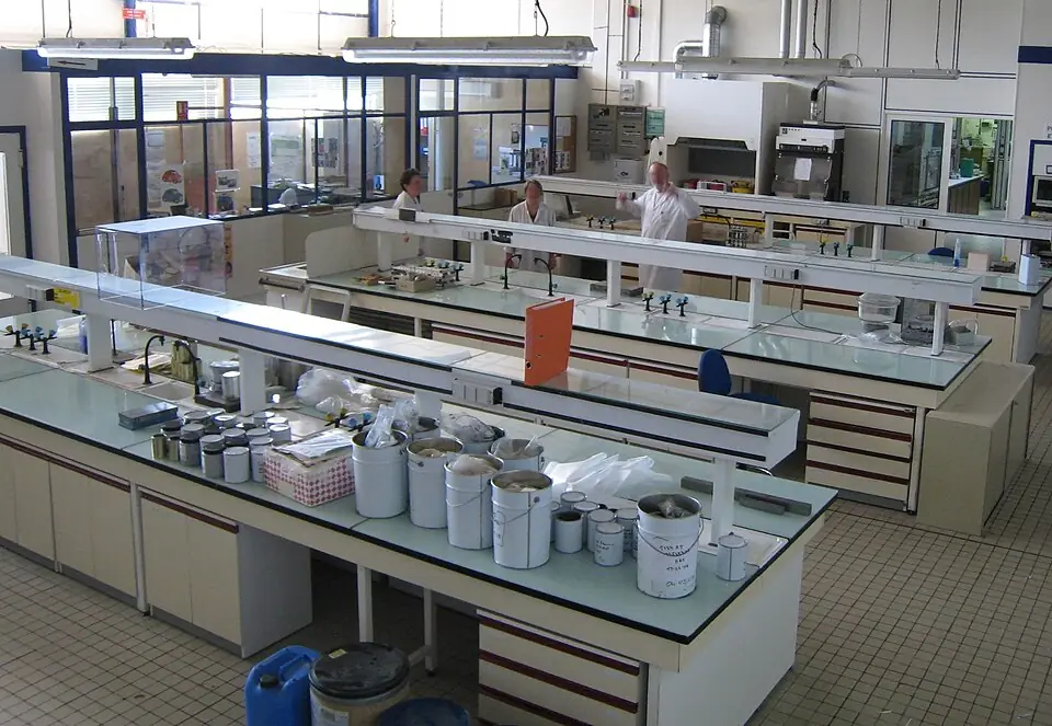
A teaching laboratory. Not a photograph of the NS&TD facility.
Cjp24, CC BY-SA 3.0. Source

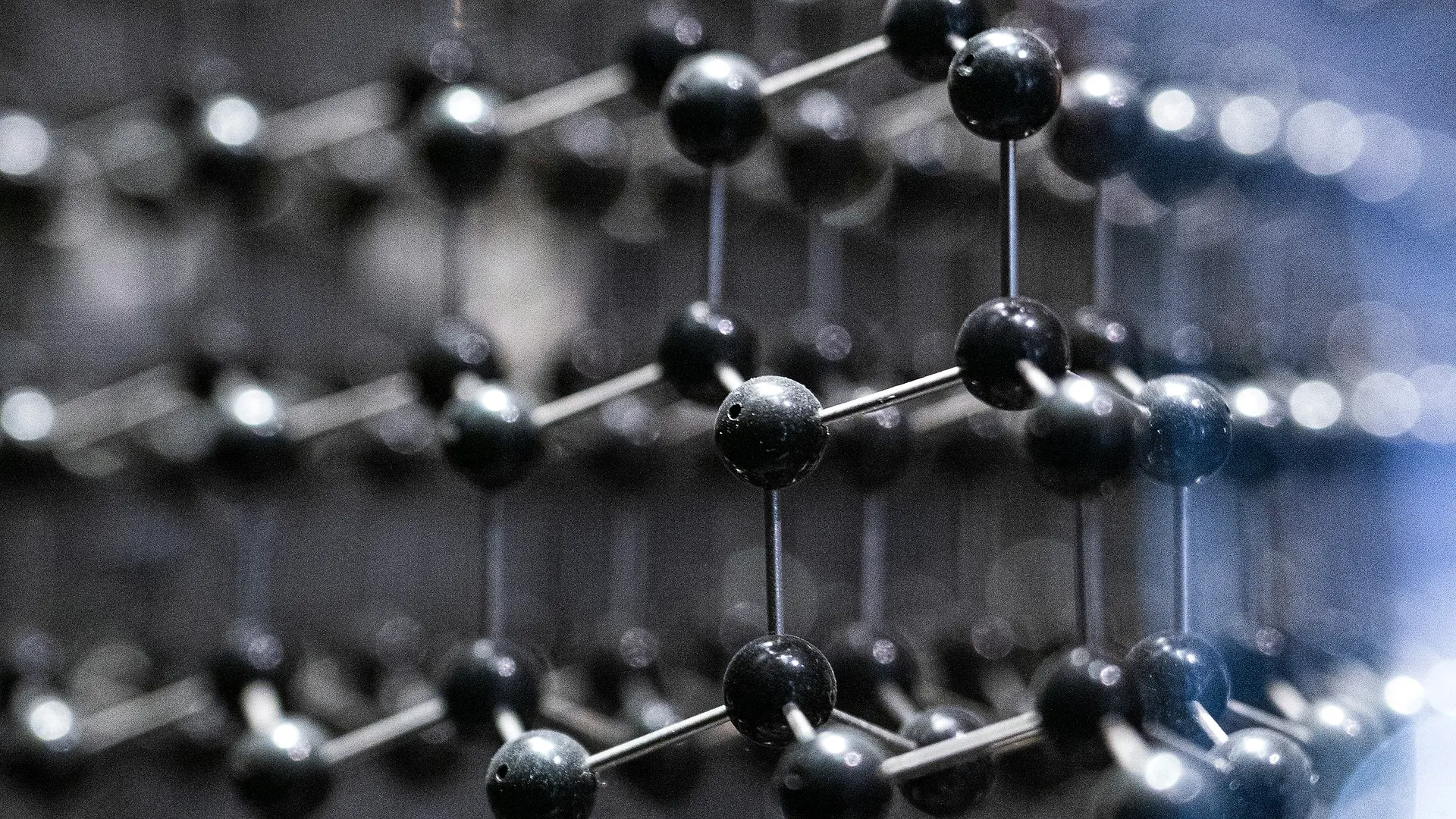
A ball-and-stick model of carbon atoms in layers, used for the carbon-materials research area.
Andy Luo, Unsplash licence. Source
Ready to submit a sample?
Download the proforma for your technique and send it with the sample, or contact the facility desk.