
National Centre for Physics · Shahdrah Valley Road, Islamabad
+92 51 2077300 Ext. 494 · nstdncp@gmail.com
Home / Instruments / FE-SEM with EDX
Microscopy & Surface Analysis

Field Emission Scanning Electron Microscopy (FE-SEM with EDX)

High-resolution imaging that uses a focused electron beam to scan a sample surface, producing detailed images of morphology and topography at the nanoscale. Coupled with energy dispersive X-ray spectroscopy (EDX), it also reports elemental composition from characteristic X-rays emitted by the sample.
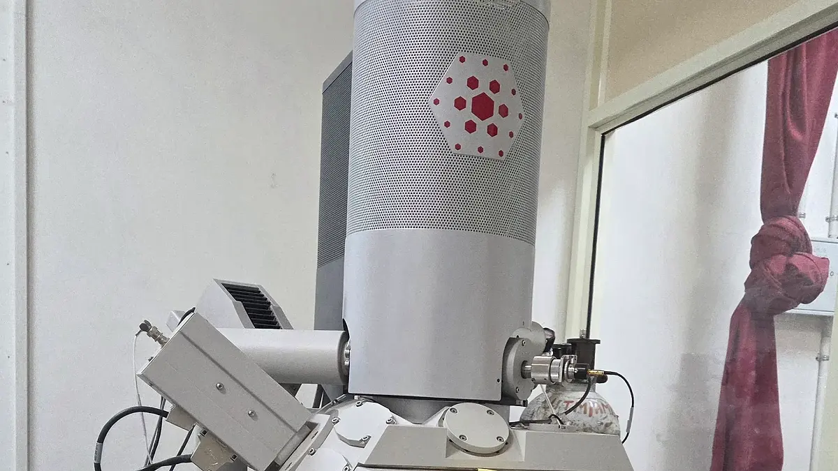
Scanning electron microscope

What the measurement provides

  • Surface morphology and topography
  • Nanoscale structural imaging
  • Elemental composition by EDX
  • Structural and chemical analysis in a single experiment

Sample requirements

  • State the nature of the sample (for example hazardous, volatile, or explosive).
  • Minimum sample quantity: 0.2 g, in an Eppendorf tube placed in a ziplock bag.
  • Only solid, powder, or thin film samples are acceptable.
  • Specify main elements for EDX analysis and expected impurities separately on the proforma.

Sample code, chemical composition, and analysis parameters for each sample.